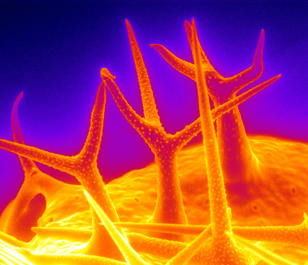
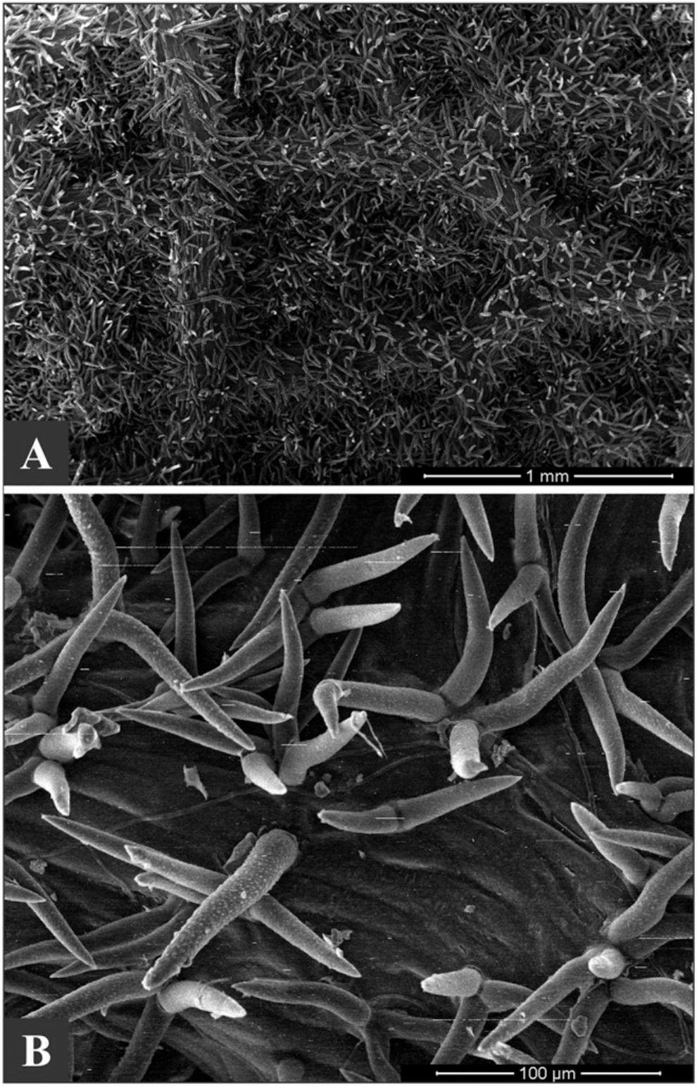
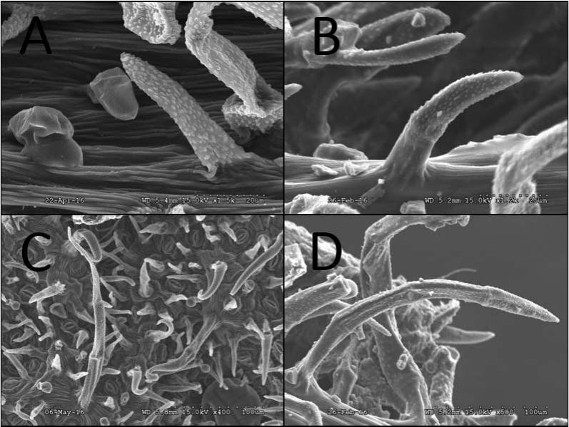
Scanning electron microscope micrographs of trichomes of tribe
Download scientific diagram | Scanning electron microscope micrographs of trichomes of tribe Neillieae. A. Neillia affinis. Capitate glandular trichome (type 4-1). B. Neillia gracilis. Distribution of long trichome (>100 µm, LS, type 1-2) at adaxial surface. C−E. Neillia sinensis var. sinensis. C. Distribution of LS (type 1-2) at abaxial surface mid vein. D, E. Unicellular LS (type 1-2) of adaxial surface. F−H. Neillia sinensis var. hypomalaca. F. Trichome surface showing smooth wall. G. Each arrows indicate short trichome (SS, type 1-1), LS (type 1-2), and subsessile glandular trichome (S, type 4-2) at abaxial surface, respectively. H. Distribution of LSs (type 1-2) at adaxial surface. I. Neillia thibetica var. thibetica. Distribution of LS (type 1-2) at abaxial surface mid vein. J. Neillia thibetica var. lobata. SS (type 1-1) and LS (type 1-2) at adaxial surface. K. Neillia thyrsiflora. S (type 4-2) at abaxial surface. L. Neillia uekii. LS (type 1-2) at adaxial surface. from publication: The systematic implication of leaf micromorphological characteristics in the tribe Neillieae (Spiraeoideae, Rosaceae) | ABSTRACT: A comparative study of the leaf epidermal micromorphology in the tribe Neillieae (Neillia: 4 species, 4 varieties; Physocarpus: 5 species; Stephanandra: 2 species) was carried out using scanning electron microscopy in order to evaluate the taxonomic and systematic | Micromorphology, Rosaceae and Leaf | ResearchGate, the professional network for scientists.

Foliar micromorphology with emphasis on the trichomes diversity

Jun-Ho SONG, Professor (Assistant), PhD, Chungbuk National University, Cheongju-si, Department of Biology

Microscopy Research and Technique, Microscopy Journal
A New Bernardia (Euphorbiaceae) with Stellate Trichomes from the

Scanning electron microscope micrographs of trichomes of tribe

A) and (B) Schematic representation of the flexural strength test. (C)
Trichome morphology relates to taxonomic diversity in Monardella

Scanning electron micrographs of the stridulatory file of Teleogryllus
Plants, Free Full-Text

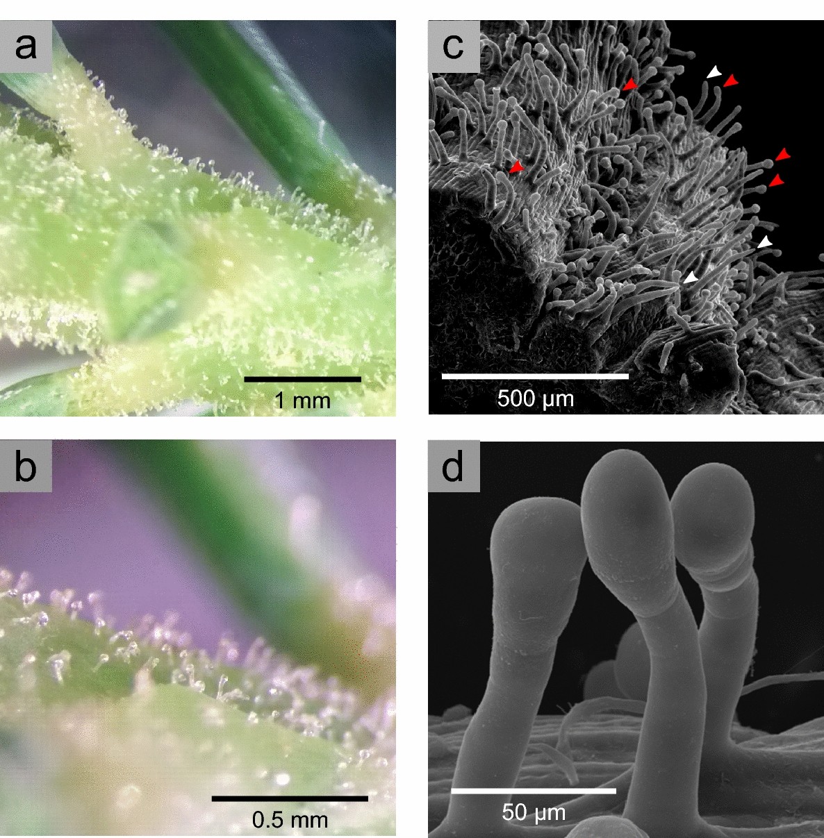
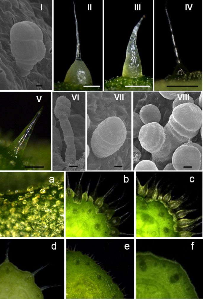
Images of glandular and non-glandular trichomes of Trichogonia
Cannabis trichomes under the electron microscope- Alchimia Grow Shop