Rose Petal Upper Surface, SEM - Stock Image - F017/4073 - Science
Papillae on the upper surface of a rose flower petal (Rosa sp), coloured scanning electron micrograph (SEM). Papillae are projections from epidermal cells and in the rose they are conical in shape. DENNIS KUNKEL MICROSCOPY/SCIENCE PHOTO LIBRARY

Universal metal printing on various surfaces. a) SEM image of rose

Focused ion beam scanning electron microscopic image of the rose petal

Compound Identification of Selected Rose Species and Cultivars: an Insight to Petal and Leaf Phenolic Profiles in: Journal of the American Society for Horticultural Science Volume 139 Issue 2 (2014)

Rose petal, SEM - Stock Image - C016/2667 - Science Photo Library

Rose Petal Upper Surface #5 Photograph by Dennis Kunkel Microscopy/science Photo Library - Pixels

Rose Petal Upper Surface, SEM - Stock Image - F017/4072 - Science Photo Library
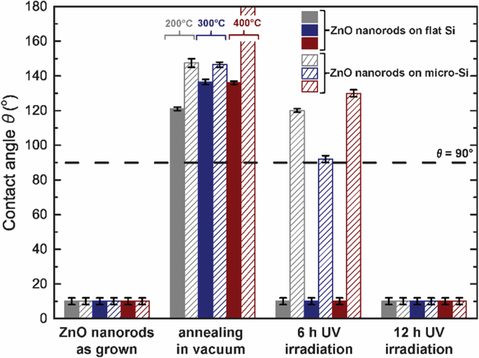
Hierarchical 'rose-petal' ZnO/Si surfaces with reversible wettability reaching complete water repellence without chemical modification

Focused ion beam scanning electron microscopic image of the rose petal
Metabolic profile and transcriptome reveal the mystery of petal blotch formation in rose, BMC Plant Biology

4 SEM images of the red rose petal surface showing the micropapillae

Rose Petal Upper Surface, SEM - Stock Image - F017/4073 - Science Photo Library

SEM of vitamin C - Stock Image - A612/0295 - Science Photo Library

Hierarchical 'rose-petal' ZnO/Si surfaces with reversible wettability reaching complete water repellence without chemical modification

Rose Petal Upper Surface #5 Photograph by Dennis Kunkel Microscopy/science Photo Library - Fine Art America

Rose petal surface. Each surface cell is 20 microns across! via @wellcomeimages